Personalized Medical Care & Dedicated International Patient Concierge

Medical Guide
Dental Implants Without Bone Grafting: 3D Imaging in Korea
Learn who qualifies for dental implants without bone grafting in Seoul, how 3D bone assessment checks volume and density, and when a graft is still required.
Schedule a ConsultationDental Implants Without Bone Grafting: Who Qualifies and How 3D Imaging Decides
Medically reviewed by Dr. Hanna Lee, DDS, MSD, PhD
Patients who have been told they need bone augmentation before receiving a replacement tooth frequently ask whether dental implants without bone graft procedures are a safe, realistic option. While promotional pages for no bone graft dental implants Korea sometimes imply that nearly anyone can skip bone augmentation through specialized techniques, clinical eligibility is strictly governed by biology and anatomy. Placing an implant directly into native jawbone—and avoiding a separate bone-grafting procedure and its healing period—is only predictable when three-dimensional diagnostics confirm adequate residual bone height, width, and density. When those structural thresholds are not met, implant placement with bone grafting remains the necessary, standard-of-care alternative.
The Bottom Line
- Implants without bone grafting place a titanium fixture directly into the patient's existing native jawbone, bypassing a separate bone-augmentation stage and its associated graft-healing period when 3D imaging verifies sufficient bone volume and density.
- Not every patient qualifies: when 3D scans reveal severe vertical or horizontal ridge resorption, thin or missing cortical walls, or insufficient bone density for primary stability, implant placement with bone grafting is the appropriate clinical alternative.
- A comprehensive 3D bone assessment before implant surgery measures three critical dimensions that flat 2D panoramic X-rays cannot fully capture: buccolingual ridge width, vertical distance to the nerve canal or sinus floor, and internal trabecular bone quality.
- At Harue Dental Clinic in Gangnam-gu, Seoul, Dr. Hanna Lee uses detailed 3D imaging, an index guided implant placement technique, and the Ankylos implant system to work precisely within eligible patients' intact native bone.
- Skipping a needed bone graft in a site that lacks adequate structural support increases the risk of poor initial fixation, thread exposure, soft-tissue recession, and early implant failure.
Why Bone Grafting Is Sometimes Recommended—and When It Can Be Avoided
To understand who qualifies for implants without bone grafting Seoul, it helps to examine why jawbone volume changes after a tooth is lost and what a dental implant needs to remain stable under daily chewing forces.
How the Jawbone Changes After Tooth Loss
Natural tooth roots are suspended in the alveolar bone by the periodontal ligament, which transmits microscopic mechanical stimuli into the surrounding jawbone every time a person chews. Once a tooth is extracted or lost to trauma or periodontal disease, that internal mechanical stimulation ceases. During the first several months after extraction, the alveolar ridge undergoes physiological remodeling and resorption, losing both horizontal width (particularly along the thinner facial or buccal bone plate) and vertical height.
In the upper back jaw, a second biological process often compounds this loss: the air-filled maxillary sinus cavity gradually expands downward into the space formerly occupied by the molar roots, a process called sinus pneumatization. Over time, these changes can leave a bony ridge that looks wide enough on the surface of the gum but is actually narrow, sloped, or shallow underneath.
When Direct Native-Bone Placement Works vs. When Grafting Is Required
A titanium dental implant requires complete bony housing around its threads and sufficient mechanical grip—known as primary stability—at the moment of insertion so that bone cells can attach to the titanium surface without micromotion during the 2–3 month osseointegration window.
- When graftless placement is appropriate: If a detailed 3D scan shows that the residual ridge still retains enough healthy width and height to surround the implant fixture—or if localized areas of bone loss or past inflammation can be bypassed by positioning the implant into an adjacent zone of dense, intact native bone—the clinician can place the implant directly without a separate bone graft.
- When bone grafting is the necessary alternative: If the ridge is too narrow to house even a narrow-diameter implant without leaving threads exposed, or if vertical bone height beneath the maxillary sinus or above the mandibular canal cannot safely accommodate a fixture, implant placement with bone grafting (either prior to or simultaneous with implant insertion) is required to rebuild the missing structural foundation.
How a 3D Bone Assessment Before Implant Surgery Decides Candidacy
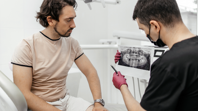
A conventional two-dimensional panoramic radiograph (orthopantomogram) flattens the curved jawbone onto a single plane. While a 2D X-ray shows approximate vertical bone height and mesiodistal space between neighboring teeth, it cannot show how thick the bone is from the cheek side to the tongue side, nor can it reveal internal hollows or localized pockets of low mineralization. That is why a 3D bone assessment before implant planning using cone-beam computed tomography (CBCT) is the decisive diagnostic step.
| Diagnostic Parameter on 3D Imaging | Favorable Finding for Graftless Implant Placement | Finding That Disqualifies Graftless Placement (Requires Bone Grafting) |
|---|---|---|
| Horizontal Ridge Width (Buccolingual) | Sufficient native bone thickness to leave intact facial and lingual cortical plates around the entire implant circumference | Knife-edge ridge or buccal wall defect where implant threads would perforate or dehisce through the bone plate |
| Vertical Bone Height (Posterior Mandible) | Adequate vertical bone height above the roof of the inferior alveolar nerve canal with a clear safety margin | Severe vertical atrophy bringing the crestal ridge too close to the mandibular nerve or mental foramen |
| Sub-Sinus Bone Height (Posterior Maxilla) | Sufficient native vertical bone beneath the maxillary sinus floor (or intact sloped sinus wall/septum suitable for targeted anchorage) | Pneumatized sinus leaving a paper-thin residual bone floor that cannot stabilize an implant without sinus augmentation |
| Trabecular Bone Density | Dense cortical shell and well-mineralized trabecular core capable of providing solid mechanical insertion torque | Severely porous or compromised bone architecture unable to provide primary mechanical fixation (low bone density implants risk) |
| Periapical and Periodontal Status | Healthy native bone free of active cysts, acute infection, or progressive vertical periodontal defects | Large unhealed cystic defects, active osteolytic infection, or widespread vertical bone loss requiring debridement and staged grafting |
Evaluating Bone Volume and Density in Three Dimensions
During the 3D evaluation at Harue Dental Clinic, the clinician inspects cross-sectional slices of the edentulous site at millimeter intervals. This allows the dentist to distinguish between two patients who might look similar on a 2D X-ray:
- Localized defect with intact basal bone: A patient may have a slight buccal concavity near the top of the ridge from an older extraction, yet 3D cross-sections reveal a broad, dense native bone column slightly deeper or palatal to the defect. Using index-guided or navigation-guided planning, the implant can be positioned entirely within that intact native bone without a separate graft.
- Knife-edge ridge requiring augmentation: Another patient may appear to have tall vertical bone on a 2D panoramic film, but the 3D cross-section reveals a ridge that is too narrow from cheek to tongue to house an implant fixture. Attempting to place an implant without grafting in that ridge would expose titanium threads; horizontal bone augmentation is essential.
Internal bone density also determines how firmly the titanium threads engage the jawbone on the day of surgery. The anterior mandible typically has thick cortical bone and dense trabecular structure, whereas the posterior maxilla naturally has thinner cortical plates and more porous bone. When planning for low bone density implants, 3D CT imaging helps the dentist evaluate cortical thickness and internal trabecular bone architecture before surgery begins. If both volume and density are inadequate to achieve initial mechanical fixation, proceeding without bone augmentation or staged healing increases the risk of micromotion and implant failure.
Who Is a Good Graftless Dental Implant Candidate—and Who Should Avoid It?
Identifying a true graftless dental implant candidate requires weighing the history of the missing tooth, the health of neighboring gums and teeth, and systemic healing factors.
Typical Profiles of Eligible Candidates
Adults who qualify for direct implant placement into native bone frequently share one or more of the following characteristics:
- Relatively recent tooth loss with preserved socket walls: Patients who lost a tooth recently without a destructive bone infection often retain most of their natural alveolar ridge dimensions before disuse atrophy sets in.
- Single missing tooth in a naturally dense jaw region: Sites in the lower jaw or premolar regions with well-preserved neighboring teeth often maintain healthy vertical bone height and thick cortical plates.
- Patients previously told they need a graft based only on 2D X-rays: Occasionally, a detailed 3D CT scan reveals an adjacent column of dense bone—such as the anterior wall of the sinus or the palatal vault of an extraction socket—that can safely house an indexed implant.
- Good general and periodontal health: Absence of active periodontal disease, uncontrolled systemic conditions (such as uncontrolled diabetes), or heavy tobacco reliance.
Clinical Findings That Disqualify a Patient from Graftless Placement
Harue Dental Clinic emphasizes that graftless implant surgery is a selective, diagnosis-driven procedure. Patients should not proceed without bone grafting when clinical and 3D evaluations identify:
- Insufficient horizontal or vertical bone volume where direct fixture placement would leave titanium threads uncovered by bone or encroach upon the inferior alveolar nerve or maxillary sinus cavity.
- Severe active periodontal bone loss or acute infection at the intended implant site, which requires periodontal therapy, infection resolution, and often regenerative grafting before an implant can be anchored.
- Long-standing edentulism with severe ridge atrophy, where years of missing teeth or removable dentures have resorbed the alveolar ridge down to basal bone lacking sufficient height.
- Compromised labial bone plate in the front-tooth zone, where placing an implant without tissue support could result in gum recession or a gray shadow showing through thin gingiva.
Can 3D Planning Help Avoid Sinus Lift Implants in the Upper Jaw?
Patients researching upper molar replacements frequently ask whether they can avoid sinus lift implants. In the posterior maxilla, if the maxillary sinus has expanded only moderately and 3D CT imaging reveals sufficient native bone height beneath the sinus floor—or sufficient bone volume just anterior or posterior to a localized sinus dip—index-guided or navigation-guided placement can position the fixture within the available native bone without opening the sinus window. When sub-sinus bone height is severely depleted across the molar span, however, sinus floor augmentation with bone grafting remains the safest way to establish vertical bone support.
Inside Harue Dental Clinic's Index-Guided Approach and Ankylos System
Located at 41-11, Gangnam-daero 162-gil, Gangnam-gu, Seoul (Sinsa-dong, Harue Dental Clinic Building), Harue Dental Clinic was established in 1995 and is led by representative director Dr. Hanna Lee. Dr. Hanna Lee graduated second in her class from Pusan National University College of Dentistry in 1994, completed her DDS, MSD, and PhD degrees in implantology at the Catholic University of Korea Graduate School of Dentistry (where she serves as an Adjunct Professor in the Department of Implant Dentistry), and completed a clinical implant surgery program at the University of Berlin. With 30+ years of experience, 90,696 cumulative implants placed (as of September 2025), and 3,000+ full-arch cases treated, Dr. Lee serves as an Asian advisor for the Ankylos implant system and a research advisor for Osstem Implant.
How Index-Guided Implant Placement Works Within Native Anatomy
When 3D imaging confirms that a patient has sufficient native bone volume and density, Dr. Lee uses an index guided implant placement technique combined with navigation-guided planning (which Harue Dental Clinic has performed since 2002 and registered as "Navigation Implant" in 2007).
Rather than making a broad surgical incision to search for bone chairside, index-guided placement references the patient's pre-mapped 3D bone coordinates and planned crown position simultaneously:
- Targeting Intact Native Bone Columns: The 3D surgical plan identifies the exact trajectory where native bone height, width, and cortical density are strongest, steering clear of localized defects or previously inflamed areas where clinically appropriate.
- Minimally Invasive Surgical Access: Because the entry point, angulation, and depth are pre-calculated from the 3D scan, the osteotomy can be prepared with minimal disruption to the surrounding periosteum and soft tissue, preserving the local blood supply that nourishes the crestal bone.
- Biomechanical Stability with the Ankylos System: Harue Dental Clinic utilizes the German-engineered Ankylos implant system, which features a progressive thread geometry and a friction-locked Morse taper conical tissue-care connection. This progressive thread design is engineered to transfer occlusal loads deeper into the trabecular bone while relieving excessive compressive stress at the delicate crestal bone margin, supporting primary stability in native bone.
How Avoiding a Separate Bone Graft Changes the Treatment Timeline
One of the practical reasons domestic and international patients inquire about graftless implant placement is the difference in overall treatment duration and surgical stages:
- Staged Bone Augmentation Pathway: When a site requires a separate pre-implant bone graft or major sinus augmentation, the grafted biomaterial often needs several months to consolidate and mineralize before the implant fixture can be placed, followed by another healing period for osseointegration.
- Direct Native-Bone (Graftless) Pathway: When 3D imaging confirms that the implant can be placed directly into sufficient native bone, the separate bone-grafting surgery and its dedicated graft-healing phase are eliminated. The implant immediately begins its standard osseointegration phase—which generally requires at least 2–3 months in immediate-loading full-arch protocols, or 2–3 primary visits across a 3–6 month window for typical single-tooth restorations (with selected high-stability front-tooth cases eligible for shortened or same-day provisional timelines).
- Coordinated In-House Restoration: Once osseointegration is verified, Dr. Lee designs a custom titanium abutment—tailored to the patient's individual gumline emergence rather than using a generic stock abutment—and fabricates the final zirconia crown in-house using the clinic's CEREC CAD/CAM digital laboratory. Prospective patients can review clinical cases in the before-and-after gallery and read verified feedback on the patient reviews page.
Travel and Communication Support for U.S. and International Patients
For patients traveling from the United States or other countries, Harue Dental Clinic is certified by the Korean Ministry of Health and Welfare as a Registered Medical Institution for Attracting Foreign Patients and operates a 19-step infection-control system. An English-speaking coordinator team (including coordinator Jeon Jin-mi) assists international visitors with reviewing recent X-rays or CT scans prior to travel, booking appointments, and managing post-treatment follow-up. Because English support is handled through the coordinator team, patients should confirm coordinator or interpreter availability for specific clinical appointments in advance via the clinic contact page.
Frequently Asked Questions
How can I find out if I qualify for dental implants without bone grafting?
Candidacy cannot be confirmed from symptoms or a visual check alone; it requires a clinical examination and a 3D cone-beam CT scan that measures the exact height, width, and density of your jawbone at the missing tooth site. If the 3D scan shows sufficient native bone volume to surround the implant and protect nearby nerves and sinuses, direct graftless placement may be an option.
What happens if my dentist tries to place an implant without a bone graft when my bone is too thin?
Proceeding without bone grafting when the existing jawbone is insufficient increases the risk of surgical and restorative complications. These include poor initial implant fixation (micromotion that prevents osseointegration), exposed titanium threads through thin facial bone, progressive gum recession around the crown, infection, or early implant failure requiring removal and staged reconstruction.
Is implant surgery without bone grafting painful?
No. Direct implant placement into native bone is performed under local anesthesia, which completely numbs the tooth site and surrounding gum tissue so that you feel only mild pressure during the procedure. Sedation options may also be available when clinically appropriate for anxious patients. Afterward, mild swelling, light bruising, or soreness for 2–3 days is common and typically manageable with prescribed medication.
How soon can international patients fly home after graftless implant placement?
When an implant is placed directly into native bone without major bone augmentation or an open sinus lift, initial recovery is often more straightforward. Many patients with uncomplicated single-implant recovery can safely fly home within 2–3 days after surgery once the dentist performs a postoperative check to confirm that initial swelling has stabilized and no bleeding or complications are present. Always confirm your exact travel schedule with the treating dentist.
Can I get a graftless implant if another clinic told me I need a bone graft?
It depends on why the bone graft was recommended and what imaging was used. If the previous recommendation was based on a 2D X-ray, a 3D CT assessment may occasionally reveal intact native bone columns where an index-guided or navigation-guided implant can be anchored without grafting. However, if the 3D scan confirms a true horizontal or vertical bone deficiency, Harue Dental Clinic will also recommend bone grafting to ensure long-term stability.
How long does the implant take to heal before the final crown is attached?
Even when bone grafting is avoided, the titanium implant fixture must still undergo biological osseointegration with your native jawbone before supporting full chewing forces on a permanent crown. Osseointegration generally takes at least 2–3 months, with many single-tooth implant cases completed across 2–3 visits over a 3–6 month period depending on bone density and site healing.
This article is for informational purposes only and is not a self-diagnosis tool or a substitute for a clinical examination and personalized treatment plan from a licensed dentist.
More Articles
Continue reading our clinical guides.