Personalized Medical Care & Dedicated International Patient Concierge

Medical Guide
Dental Implants With vs Without Bone Grafting Compared
Compare dental implants with or without a bone graft, including 3D scan bone density rules, healing timelines, and suitability in Seoul, South Korea.
Schedule a ConsultationDental Implants With vs Without Bone Grafting: Timeline, Healing and Suitability Compared
Medically reviewed by Dr. Hanna Lee, DDS, MSD, PhD
The Bottom Line
- Choosing between dental implants with or without bone graft procedures is a diagnostic decision driven by three-dimensional jawbone measurements—specifically residual bone height, ridge width, and trabecular density—rather than a patient or marketing preference.
- At Harue Dental Clinic in Gangnam-gu, Seoul, eligible patients whose 3D imaging confirms adequate native bone volume and density can receive direct implant placement without a separate bone-augmentation procedure, avoiding the additional graft-healing phase.
- Navigation-guided 3D planning (used at the clinic since 2002 and registered as "Navigation Implant" with the Korean Intellectual Property Office in 2007) maps nerve pathways, sinus anatomy, and existing bone architecture to identify whether compromised or inflamed zones can be bypassed safely.
- When 3D assessment reveals insufficient bone volume or density to achieve predictable primary implant stability, bone grafting—either performed prior to implantation or combined with placement—remains the essential clinical alternative, because omitting necessary augmentation elevates the risk of inadequate fixation and implant failure.
- For international travelers visiting Seoul, understanding whether your anatomy supports a graftless route or requires staged bone augmentation is the single largest factor determining how many trips you will make and how long you must wait before receiving your definitive zirconia crown.
Why the Question "Do I Need a Bone Graft for a Dental Implant?" Matters So Much
When a natural tooth is extracted or lost to trauma, fracture, or periodontal disease, the surrounding alveolar bone no longer receives the mechanical chewing forces that stimulate continuous bone remodeling. Over the months and years following tooth loss, the jawbone naturally resorbs in both horizontal width and vertical height. For patients researching tooth replacement—especially U.S. patients planning care in South Korea—one of the first and most consequential clinical questions is: do I need a bone graft for a dental implant, or can a titanium fixture be anchored directly into the bone I already have?
The answer shapes every practical aspect of your treatment journey:
- Surgical Complexity and Morbidity: Direct placement into native bone involves a single surgical site where the implant fixture is inserted. Adding a separate bone-augmentation or sinus-lifting step introduces additional biomaterials, membrane stabilization, and broader soft-tissue management.
- Total Treatment Timeline: While standard osseointegration of an implant in native bone requires a dedicated healing window before final restoration, a staged bone graft adds a separate graft-maturation interval before the implant can even be placed—or extends the integration window when grafting and placement are performed simultaneously.
- Travel Logistics for Overseas Patients: For patients flying from Los Angeles, New York, San Francisco, Houston, or Chicago to Seoul, avoiding an unnecessary separate grafting stage can eliminate an entire transpacific trip or shorten the interval between initial surgery and final prosthetic restoration.
At the same time, attempting a graftless implant when the residual ridge is genuinely deficient is a serious clinical hazard. If a titanium fixture is placed into bone that lacks sufficient volume or density to lock the threads in place, micro-movement during early healing can prevent osseointegration, lead to soft-tissue recession, or result in implant loss. The goal of modern digital implantology is never to force a graftless procedure onto unsuitable anatomy, nor to perform routine bone grafting by default when healthy native bone is already available. Instead, detailed 3D diagnostics allow the clinician to match the pathway to the patient's exact anatomy.
Understanding the Two Pathways: Bone Grafting vs Graftless Implants
To compare bone grafting vs graftless implants clearly, it helps to examine what happens biologically and mechanically in each approach.
Pathway 1: Implants Without Bone Grafting (Direct Native-Bone Placement)
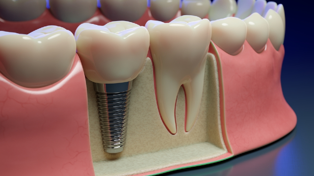
In an Implant Without Bone Grafting procedure, a titanium implant fixture is placed directly into the patient's existing jawbone without a separate bone-grafting or bone-augmentation surgery. This pathway is viable when detailed 3D imaging confirms that the native alveolar ridge possesses enough vertical height, buccolingual width, and mineral density to surround the fixture and provide strong mechanical primary stability on the day of surgery.
At Harue Dental Clinic, Director Dr. Hanna Lee—who has 30+ years of clinical experience in implantology and aesthetic dentistry, graduated 2nd in her class from Pusan National University College of Dentistry in 1994, serves as an Adjunct Professor in the Department of Implant Dentistry at the Graduate School of Dentistry at The Catholic University of Korea, and completed a clinical implant surgery program at the University of Berlin—evaluates each implant site using 3D imaging and an index-guided, minimally invasive surgical technique. Using the Ankylos implant system and computer-assisted navigation guidance, the treatment plan evaluates whether compromised or inflamed areas of bone can be avoided by selecting an optimal angle, depth, and position within healthy native bone.
When the patient's existing bone structure is sufficient, direct placement offers several distinct advantages:
- It avoids a separate bone-augmentation surgery and the biological healing period required for graft consolidation.
- It relies entirely on the patient's vital, vascularized native bone for osseointegration.
- It generally shortens the overall treatment timeline from initial consultation to definitive crown placement compared with staged augmentation pathways.
Pathway 2: Dental Implants With Bone Grafting (Simultaneous or Staged Augmentation)
When clinical evaluation and 3D imaging demonstrate that the existing jawbone cannot stably house an implant fixture on its own, Implant With Bone Grafting serves as the established restorative pathway. Bone deficiency typically arises from long-term tooth loss, advanced periodontal bone loss, trauma during a previous tooth extraction, or natural pneumatization (expansion) of the maxillary sinus in the upper back jaw.
Depending on how much native bone remains, graft-assisted implant therapy generally follows one of two schedules:
- Simultaneous Augmentation and Implant Placement: When the residual bone is slightly narrow or has a localized defect—yet still retains enough basal bone to lock the implant fixture with adequate initial stability—the dentist can place the titanium implant and pack bone graft material (often covered by a barrier membrane in guided bone regeneration, or GBR) during the same surgical session. This avoids a second surgical appointment for implant placement, though the combined site often requires a longer healing period before the definitive crown is loaded.
- Staged Bone Grafting Followed by Delayed Implantation: When bone loss is severe and the residual ridge cannot provide mechanical primary stability for an implant fixture, the bone graft must be performed first as a standalone procedure. Only after the grafted site has healed and matured into a stable osseous foundation is the dental implant surgically placed in a subsequent visit.
Side-by-Side Comparison: Graftless vs Graft-Assisted Implant Pathways
The table below outlines how direct native-bone placement compares with simultaneous and staged bone-grafting pathways across diagnostic criteria, surgical steps, healing biology, and travel planning.
| Clinical Dimension | Implants Without Bone Grafting (Graftless) | Implants With Simultaneous Bone Graft | Implants With Staged Bone Graft (Delayed Placement) |
|---|---|---|---|
| Primary Anatomical Requirement | Sufficient native bone height, width, and density confirmed on 3D imaging to house the fixture with strong primary stability | Enough native bone to stabilize the implant fixture initially, but localized contour or volume deficiency requiring augmentation | Insufficient residual bone volume or density to achieve mechanical primary stability at the time of initial surgery |
| Typical Clinical Scenarios | Recent tooth extraction with intact socket walls; single missing tooth in naturally dense bone; sites where navigation planning can bypass minor defects | Moderate horizontal ridge thinning; minor dehiscence or fenestration around an otherwise stable implant; moderate sinus floor proximity | Severe long-term ridge resorption; extensive periodontal bone destruction; severely pneumatized maxillary sinus with minimal residual bone height |
| Number of Surgical Stages | Single surgical placement stage directly into native jawbone | Single combined surgical stage (implant placement plus concurrent bone augmentation) | Two distinct surgical stages: first bone augmentation, followed months later by implant fixture placement |
| Biological Healing Process | Direct osseointegration between vital native host bone and the titanium implant surface | Concurrent graft vascularization/remodeling and bone-to-implant osseointegration | Phase 1: Graft maturation and remodeling into vital bone; Phase 2: Implant osseointegration after delayed placement |
| Overall Treatment Timeline | Shortest overall pathway; avoids a separate graft-healing phase prior to restoration | Moderate timeline; osseointegration period is often extended compared with native-bone placement to allow graft maturation | Longest overall pathway; requires a separate graft-healing window before the implant osseointegration timeline even begins |
| International Travel Impact | Often completed in 2–3 visits over 3–6 months for single/front teeth, or 4 visits over 4–6 months for eligible full-arch cases | Usually follows a two-trip structure (surgery trip plus restorative trip), with a slightly longer interval between trips | Frequently requires three separate trips to Seoul (grafting trip, implant placement trip, and final crown/prosthesis trip) |
How 3D Scan Bone Density and Anatomy Decide the Route
A common frustration among dental patients is receiving conflicting opinions from different clinics—one dentist stating that a bone graft is mandatory, and another suggesting that implants can be placed without grafting. Why do clinical opinions diverge, and how should a patient evaluate them?
Limitations of Two-Dimensional Panoramic X-Rays
Standard two-dimensional panoramic radiographs flatten complex three-dimensional jaw anatomy into a single plane. While a panoramic X-ray shows approximate vertical bone height and mesiodistal space between tooth roots, it cannot accurately measure buccolingual ridge width (how thick the jawbone is from the cheek side to the tongue side) or evaluate internal trabecular bone density. A ridge that looks tall enough on a flat X-ray may actually be knife-edged and too thin to surround an implant neck, or conversely, a site that looks questionable on a 2D film may possess dense, well-preserved bone just a few millimeters away at a slightly tilted trajectory.
What 3D Imaging Reveals Before Surgery
At Harue Dental Clinic, every implant consultation includes a detailed 3D imaging assessment of the surgical site. Rather than relying on freehand visual estimation during surgery, Dr. Hanna Lee—who has placed 90,696 cumulative dental implants as of September 2025 and treated 3,000+ full-arch cases—uses 3D CT and optical camera scanning to evaluate four specific anatomical variables:
- Vertical Bone Height to Vital Structures: In the upper posterior jaw, 3D imaging maps the exact floor of the maxillary sinus. In the lower posterior jaw, it traces the path of the inferior alveolar nerve canal. Knowing the exact millimeter clearance prevents nerve injury or unintended sinus perforation.
- Horizontal Ridge Width and Cortical Plate Integrity: The scan shows whether the facial (outer) and lingual/palatal (inner) bone plates are intact. In front-tooth implant cases, preserving and supporting the labial bone plate is essential for maintaining natural gumline contour and preventing soft-tissue shadow or recession.
- Trabecular Bone Density: Bone is not uniform throughout the mouth. The anterior mandible typically has dense cortical and trabecular architecture, whereas the posterior maxilla often consists of softer, more porous cancellous bone. Evaluating 3D scan bone density helps determine whether an implant will achieve the initial mechanical torque required for immediate or early stabilization.
- Compromised or Inflamed Zones: When a tooth has failed due to endodontic infection or localized periodontal disease, parts of the socket may be resorbed or inflamed. Navigation-guided 3D planning allows the dentist to map the surrounding healthy bone and determine whether the implant trajectory can avoid compromised areas—minimizing or potentially avoiding extensive bone grafting where clinically appropriate.
Sinus Lift vs Graftless Approaches in the Upper Back Jaw
The posterior maxilla (upper molar and premolar region) is the area of the mouth most frequently associated with bone grafting questions. After upper back teeth are lost, the bone ridge resorbs from below while the air-filled maxillary sinus cavity expands downward from above, often leaving a thin bridge of bone between the oral cavity and the sinus floor.
When patients compare sinus lift vs graftless options, 3D surgical planning evaluates three distinct clinical possibilities depending on the remaining anatomy:
1. Navigation-Guided Placement Within Available Native Bone
If 3D imaging demonstrates that sufficient native bone volume exists—either directly beneath the sinus floor or by angling the implant fixture into adjacent dense bone columns (such as the anterior sinus wall or tuberosity/pterygoid region in full-arch configurations)—the implant can be placed using real-time navigation guidance without breaching the sinus membrane or performing a separate sinus graft.
2. Targeted Transcrestal or Simultaneous Sinus Elevation
When vertical bone height is moderately reduced but still offers enough native bone thickness to stabilize the implant neck, the clinician may elevate the sinus membrane conservatively through the implant osteotomy site, with or without localized graft material depending on the defect geometry and clinical protocol. Peer-reviewed systematic reviews show that when primary implant stability is achieved, simultaneous approaches provide high survival rates while sparing the patient a separate precursor surgery.
3. Lateral Window or Staged Sinus Augmentation
When the residual bone beneath the maxillary sinus is too thin to hold an implant fixture stably, placing an implant without augmentation risks displacement into the sinus cavity or early failure. In these low-bone cases, building vertical bone height through sinus floor augmentation is the necessary foundation before or alongside implant placement.
Dental Implant Bone Graft Healing Time vs Native-Bone Osseointegration
Patients comparing dental implant bone graft healing time with graftless recovery should distinguish between two different biological clocks: short-term soft-tissue recovery (the first few days after surgery) and deep osseous healing (the months required for bone remodeling and osseointegration).
Immediate Post-Surgical Recovery (Days 1 to 7)
Regardless of whether bone grafting is performed, dental implant placement is conducted under local anesthesia (with sedation options available when clinically appropriate to help with anxiety or relaxation), so patients do not feel sharp pain during the procedure itself.
- After Graftless Implant Placement: Because no separate donor site or extensive periosteal flap release for graft containment is needed, initial post-surgical downtime is typically compact. Harue Dental Clinic documents that mild swelling, light bruising, or slight soreness for 2 to 3 days is normal and manageable with prescribed medication. Many international patients with uncomplicated graftless or single front-tooth implant placement can safely fly home 2 to 3 days after surgery once initial swelling stabilizes and the dentist completes a post-operative check.
- After Implant Placement With Bone Grafting or Sinus Lifting: When bone augmentation or sinus floor elevation is performed, patients may experience somewhat more pronounced facial swelling, bruising, or sinus-area pressure during the first week. Flying home may require a slightly longer buffer in Seoul so the dentist can verify stable wound closure and absence of bleeding or sinus complications before cabin pressure exposure.
Deep Bone Integration and Overall Implant Treatment Time With Graft vs Without
Once the gums heal on the surface, the underlying bone must lock onto the microscopic surface of the titanium implant fixture—a process called osseointegration.
- Graftless Single-Tooth and Front-Tooth Timeline: When an implant is placed directly into adequate native bone, no separate graft-consolidation window is needed. At Harue Dental Clinic, front-tooth implant treatment typically requires 2 to 3 primary visits across a 3 to 6 month window, with the surgical placement session taking approximately 60 to 90 minutes. In selected front-tooth cases where bone density and site conditions are ideal, a shortened restorative timeline or same-day implant and provisional prosthetic treatment may be considered after individual evaluation.
- Graftless Immediate-Loading Full-Arch Timeline: For patients undergoing full-arch restoration (such as All-on-4 or All-on-6) with sufficient strategic bone support and initial stability, Harue Dental Clinic connects a temporary fixed prosthesis on the same day as surgery using a bilateral load-balance protocol. Osseointegration and guided bone healing take approximately 2–3 months (during which patients must avoid excessive biting force on the temporary teeth), and documented full-arch cases are typically completed in 4 visits over 4–6 months.
- Implant Treatment Time With Graft: When bone grafting is required, the biological timeline expands because grafted particles must first be infiltrated by host blood vessels and gradually replaced by vital, load-bearing bone. If grafting is performed simultaneously with implant placement, the osseointegration waiting period before fitting the definitive zirconia crown is commonly extended toward the upper end of the 3 to 6 month range (or longer depending on defect size). If severe bone loss requires a staged bone graft prior to implant placement, the patient must first complete several months of graft maturation before the implant fixture is placed, followed by an additional osseointegration period—extending total treatment across multiple quarters.
Assessing Implant Candidacy in Low Bone: Who Qualifies for Each Route?
Determining implant candidacy low bone status is never a matter of guesswork. During an in-person evaluation at Harue Dental Clinic, suitability for graftless versus graft-assisted treatment is organized around clear clinical benchmarks.
Who Is a Strong Candidate for Implants Without Bone Grafting?
You are more likely to qualify for direct native-bone implant placement if:
- You lost the tooth relatively recently, before significant horizontal or vertical alveolar ridge resorption has occurred.
- The missing tooth is in a naturally dense anatomical zone, or 3D imaging confirms sufficient bone height, width, and density at the intended implant site.
- Surrounding periodontal tissues are healthy, without active gum infection or widespread periodontal bone loss in the treatment area.
- Strategic 3D angulation can utilize adjacent bone, such as in All-on-4 or All-on-6 full-arch configurations where four or six implants are positioned strategically across the arch to maximize contact with available native bone rather than placing an individual implant into every resorbed posterior tooth socket.
Who Requires Bone Grafting or Staged Preparation First?
A graftless approach is not suitable—and bone augmentation or delayed implantation is recommended instead—when:
- 3D imaging shows insufficient bone volume or density to surround the implant fixture or achieve adequate mechanical primary stability.
- Active infection or severe inflammation has destroyed the socket walls, requiring debridement and site regeneration before an implant can be safely anchored.
- Anterior aesthetic architecture is compromised: In the upper front teeth, if the thin facial bone plate has resorbed following trauma or long-term tooth loss, placing an implant without hard- or soft-tissue support can result in an unnatural crown length or visible gumline recession.
- Systemic or lifestyle risk factors are uncontrolled: Regardless of whether a graft is used, patients with severe uncontrolled diabetes, untreated active periodontal disease, or heavy tobacco use face elevated risks of delayed healing, infection, and osseointegration failure.
How Harue Dental Clinic Coordinates Surgical Placement and In-House Crown Design
Whether your 3D scan directs you toward a graftless implant or a graft-assisted pathway, long-term functional and aesthetic success depends on how well the surgical implant position matches the final crown. If an implant is placed purely where bone happens to be thickest—without regard for how the replacement tooth will meet the opposing bite—the resulting crown can suffer from off-axis chewing forces, food trapping, or an unnatural emergence profile.
At Harue Dental Clinic, surgical placement and prosthetic restoration are never treated as disconnected steps handled by separate providers:
- Single-Doctor Continuity: Dr. Hanna Lee personally oversees every stage of care, from initial 3D diagnostic mapping and navigation-guided surgical placement to custom abutment design and final crown fitting.
- Prosthetically Driven Index-Guided Placement: Using 3D camera imaging and navigation software, the intended final crown shape, gumline contour, tooth proportion, and bite alignment are planned before surgery begins. During placement of the Ankylos implant fixture, an index-guided technique aligns the implant angle and depth directly with the planned restoration.
- Custom Titanium Abutments and In-House Zirconia Crowns: Once osseointegration is confirmed, Harue Dental Clinic designs a custom titanium abutment tailored to the individual implant site and gum margin rather than relying on a one-size-fits-all stock abutment. The definitive metal-free zirconia crown is then digitally designed and milled in the clinic's in-house CEREC digital laboratory (established in 2007), allowing chairside refinement of shade, translucency, and bite comfort. You can review documented clinical cases in the clinic's before-and-after gallery and read international patient experiences on the reviews page.
- Rigorous Infection Control: Every surgical and restorative visit is supported by the clinic's 19-step infection-control and sterilization system at its facility in Gangnam-gu, Seoul (41-11, Gangnam-daero 162-gil, Sinsa-dong, Harue Dental Clinic Building).
Practical Planning Checklist for International Patients
If you are traveling to South Korea from the United States and want to know how bone grafting possibilities affect your itinerary, use the following step-by-step framework before booking flights:
- Step 1: Send Existing Dental Records Before You Fly. Reach out to Harue Dental Clinic's English-speaking coordinator team (led by coordinator Jeon Jin-mi) via the online contact page, phone (
+82 2 543 0037), WhatsApp (+1 208-825-7756), or email (info@haruedentalclinic.com). Share any recent panoramic X-rays or 3D CBCT scans, along with your medical history and current medication list. - Step 2: Treat Remote Reviews as Preliminary. While pre-travel imaging helps the team estimate whether you may be a candidate for graftless placement, immediate loading, or bone augmentation, final suitability and your exact treatment timeline can only be confirmed after an in-person clinical examination and 3D scan at the clinic in Seoul.
- Step 3: Build Buffer Days Into Your Initial Trip. If your case is straightforward and qualifies for implants without bone grafting, initial recovery is typically rapid, with many patients able to fly home within 2 to 3 days once swelling stabilizes and the post-op check is complete. If full-arch All-on-4 treatment is planned, patients typically stay around 7–14 days for consultation, surgery, same-day temporary teeth, and initial follow-up. If bone grafting or sinus elevation is possible, allow flexible post-operative days in Seoul so the dentist can confirm safe travel clearance.
- Step 4: Plan for the Integration Interval. Whether your osseointegration period is approximately 2–3 months (for immediate-loading full-arch cases) or a 3 to 6 month window (for staged single or front-tooth implants), coordinate the timing of your return restorative trip with the coordinator team before leaving Seoul.
Frequently Asked Questions
How do I know for sure if I need a bone graft for a dental implant?
You cannot know with certainty from a visual exam or a flat 2D X-ray alone. Definitive determination requires a 3D imaging scan that measures bone height, buccolingual width, and mineral density in three dimensions, alongside a clinical exam of your gums and adjacent teeth. If the 3D scan confirms sufficient native bone volume and density to stabilize the implant fixture safely, direct placement without bone grafting may be performed; if bone support is inadequate, bone augmentation is recommended.
What happens if a dentist places an implant without a bone graft when my bone is actually too thin?
Proceeding with direct implant placement when the existing jawbone is insufficient significantly increases the risk of surgical and biological complications. Without adequate bone thickness and density around the titanium threads, the implant may fail to achieve initial mechanical fixation, suffer from micro-movement during early healing, develop gum recession that exposes the metal collar, or fail to osseointegrate altogether—ultimately requiring implant removal and revision surgery.
Does navigation-guided implant surgery eliminate the need for bone grafting in every patient?
No. Navigation-guided implant surgery uses computer-assisted 3D mapping to control the angle, depth, and position of the implant fixture with high precision, which can help the dentist utilize existing healthy bone and avoid compromised or inflamed areas where clinically appropriate. While this allows selected borderline cases to avoid or minimize extensive bone grafting, patients who lack the minimum foundational bone volume required to anchor an implant still need bone augmentation.
Is recovery after implants without bone grafting faster than recovery with a bone graft?
Yes, in terms of both initial post-operative comfort and total time to final restoration. Without a separate bone-augmentation procedure, initial surgical trauma is more localized—typically involving mild swelling, light bruising, or slight soreness for 2 to 3 days—and you avoid the additional months required for a staged bone graft to mature before implant integration can finish. However, the implant fixture itself still requires a biological osseointegration period before the definitive crown is placed.
Can I still get an implant without bone grafting if another dentist told me I needed a graft?
It depends on why the graft was recommended and what type of imaging was used. If the previous recommendation was based only on a 2D X-ray or freehand estimation, a detailed 3D scan and navigation-guided assessment at Harue Dental Clinic can clarify whether sufficient native bone exists along an alternative angulation or whether a strategic configuration (such as All-on-4 or All-on-6 for full-arch cases) can utilize denser anterior bone zones. If the 3D scan confirms true bone deficiency at the required site, however, bone grafting will still be advised.
When is it safe to fly home after dental implant surgery with or without bone grafting?
After uncomplicated implant placement without bone grafting, many international patients can safely fly home within 2 to 3 days once initial swelling stabilizes and the dentist completes a post-operative check to confirm there is no active bleeding or complication. When bone grafting, sinus lifting, or full-arch surgery (which typically involves a 7–14 day stay in Korea) is performed, travel timing depends on the extent of augmentation and your individual recovery, and must always be confirmed directly with your treating dentist.
This article is for informational purposes only and is not a self-diagnosis tool or a substitute for a clinical examination and personalized treatment plan from a licensed dentist.
More Articles
Continue reading our clinical guides.